Anatomy Of Chest / Knowledge Assessment : Related posts of anatomy of the chest area abdominal muscles picture anatomy.. The dominant muscle in the upper chest is the pectoralis major. This chapter is an abbreviated review of thoracic anatomy as seen on chest radiographs and computed tomography (ct) of the chest. Radiology basics of chest ct anatomy with annotated coronal images and scrollable axial images to help medical students and junior doctors learning anatomy. The chest or thorax region of the upper body has a number of important organs that reside within it that may present with chest pain if they become compromised in. It is enclosed by the ribs, the vertebral column, and the sternum, or breastbone, and is separated from the abdominal cavity (the body's largest hollow space) by a muscular and membranous partition, the diaphragm.
Thoracic cavity, also called chest cavity, the second largest hollow space of the body. See human chest anatomy stock video clips. Anatomy of the thorax, heart, abdomen and pelvis recommended text gray's anatomy for students. Learn about each of these muscles, their locations, functional anatomy and exercises for them. Radiology basics of chest ct anatomy with annotated coronal images and scrollable axial images to help medical students and junior doctors learning anatomy.
Pectus Carinatum - Causes, Symptoms, Brace & Surgery Treatment from healthjade.com 1 effects 2 notes 3 trivia 4 see also improves the contents of broken chests. Anatomically, the heart is located in the anterior thoracic cavity; The superior thoracic aperture found superiorly and the inferior thoracic aperture. The chest is the area of origin for many of the body's systems as it houses organs such as the heart, esophagus, trachea, lungs, and thoracic diaphragm. Abdominal muscles picture anatomy 12 photos of the abdominal muscles picture anatomy abdominal muscles anatomy diagram, abdominal muscles picture anatomy, human anatomy, abdominal muscles anatomy diagram, abdominal muscles picture anatomy Learn about each of these muscles, their locations, functional anatomy and exercises for them. Computed tomography (ct) of the chest can detect pathology that may not show up on a conventional chest radiograph(1). Plus, how to target each to make them bigger and stronger.
Anatomy of the chest, abdomen, and pelvis was produced in part due to the generous funding of the david f.
Angina is the term for chest pain caused by poor blood flow to the heart. The pectoralis major and the pectoralis minor, known collectively as your pecs. It provides protection to vital organs (eg, heart and major vessels, lungs, liver) and provides stability for movement. It is important to remember the position and orientation of the heart when placing a stethoscope on the chest of a patient and listening for heart sounds, and also when looking at images taken from a midsagittal perspective. Chest pain has many possible causes, all of which need medical attention. The superior thoracic aperture found superiorly and the inferior thoracic aperture. The epidermis is the outermost layer that provides a protective, waterproof seal over the body. Anatomy of right side chest pain. Anatomy of the thorax, heart, abdomen and pelvis recommended text gray's anatomy for students. See human chest anatomy stock video clips. The thorax has two major openings: It removes junk, and explosions from the destruction table while replacing them with pickups (such as , , , etc), it also improves the quality of dropped items. A good radiologist knows the anatomy because knowing where structures normally live and recognizing the location of an abnormality helps to make or narrow the differential diagnosis.
It is enclosed by the ribs, the vertebral column, and the sternum, or breastbone, and is separated from the abdominal cavity (the body's largest hollow space) by a muscular and membranous partition, the diaphragm. #anatomy of the chest and stomach. Anatomy of the chest and the lungs: Thoracic cavity, also called chest cavity, the second largest hollow space of the body. Anatomy of the thorax, heart, abdomen and pelvis recommended text gray's anatomy for students.
01 CXR Anatomy - YouTube from i.ytimg.com (1) the pectoralis major, and (2) the pectoralis minor. A good radiologist knows the anatomy because knowing where structures normally live and recognizing the location of an abnormality helps to make or narrow the differential diagnosis. Anatomically, the heart is located in the anterior thoracic cavity; Applied anatomy of the chest wall and mediastinum petros mirilas michael e. Related posts of anatomy of the chest area abdominal muscles picture anatomy. Skandalakis chest wall embryogenesis the muscles of the chest develop from the somites found in the mesoderm. Anatomy of the chest and stomach, human anatomy, anatomy of the chest and stomach. Anatomy of the chest and the lungs:
Applied anatomy of the chest wall and mediastinum petros mirilas michael e.
Thoracic cavity, also called chest cavity, the second largest hollow space of the body. The first step in understanding thorax anatomy is to find out its boundaries. The chest is made up primarily of two muscles: The chest is the area of origin for many of the body's systems as it houses organs such as the heart, esophagus, trachea, lungs, and thoracic diaphragm. Browse 6,407 chest anatomy stock photos and images available, or search for human anatomy to find more great stock photos and pictures. Here, we break down the anatomy of your chest muscles. It provides protection to vital organs (eg, heart and major vessels, lungs, liver) and provides stability for movement. Anatomy of the chest and stomach, human anatomy, anatomy of the chest and stomach. Learn about each of these muscles, their locations, functional anatomy and exercises for them. The chest or thorax region of the upper body has a number of important organs that reside within it that may present with chest pain if they become compromised in. The dominant muscle in the upper chest is the pectoralis major. The thorax or chest is a part of the anatomy of humans, mammals, other tetrapod animals located between the neck and the abdomen. Anatomy of the thorax, heart, abdomen and pelvis recommended text gray's anatomy for students.
Related posts of anatomy of the chest area abdominal muscles picture anatomy. The pectoralis major and the pectoralis minor, known collectively as your pecs. (1) the pectoralis major, and (2) the pectoralis minor. Book of chest anatomy is a passive item. Learn about each of these muscles, their locations, functional anatomy and exercises for them.
Chest anatomy, artwork - Stock Image - F006/1230 - Science ... from media.sciencephoto.com Chest bone, ribs, lung, heart, xiphoid process, sternum anatomy. The first step in understanding thorax anatomy is to find out its boundaries. The dominant muscle in the upper chest is the pectoralis major. Related posts of anatomy of the chest area abdominal muscles picture anatomy. Here, we break down the anatomy of your chest muscles. Anatomy of the chest and the lungs: The thorax or chest is a part of the anatomy of humans, mammals, other tetrapod animals located between the neck and the abdomen. This chapter is an abbreviated review of thoracic anatomy as seen on chest radiographs and computed tomography (ct) of the chest.
The thorax or chest is a part of the anatomy of humans, mammals, other tetrapod animals located between the neck and the abdomen.
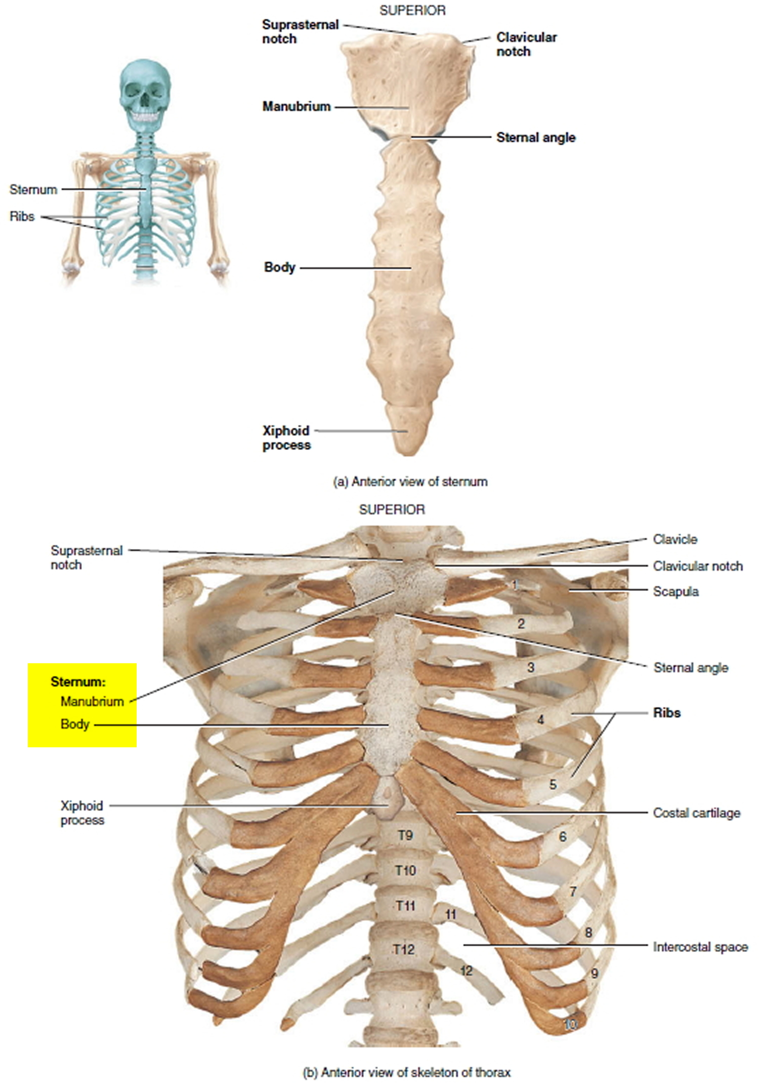
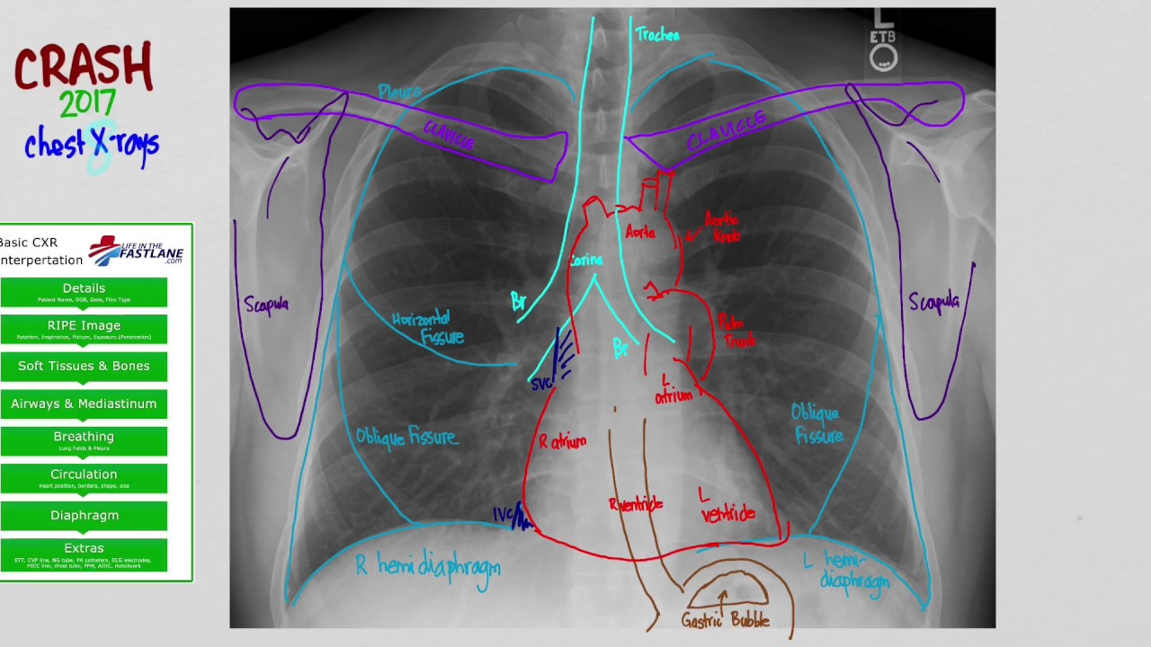
Book of chest anatomy is a passive item. Thoracic cavity, also called chest cavity, the second largest hollow space of the body. This page provides an overview of the chest muscle group. Plus, how to target each to make them bigger and stronger. The chest wall is comprised of skin, fat, muscles, and the thoracic skeleton. Chest bone, ribs, lung, heart, xiphoid process, sternum anatomy. In insects, crustaceans, and the extinct trilobites, the thorax is one of the three main divisions of the creature's body, each of which is in turn composed of multiple segments. The chest or thorax is the region between the neck and diaphragm that encloses organs, such as the heart, lungs, esophagus, trachea, and thoracic diaphragm. Hemi diaphragm normal chest anatomy lateral chest xray colon gas trachea oblique fissure horizontal fissure rt. It provides protection to vital organs (eg, heart and major vessels, lungs, liver) and provides stability for movement. In this image, you will find common carotid arteries, internal jugular vein, subclavian artery, subclavian vein, heart, right lung, 6th rib, diaphragm, costal cartilage in it. You will also find the xiphoid process, 10th rib, the apex of the heart, the coronary vein of the heart. Sternocleidomastoid muscle clavicle and ribs anatomy muscle anatomy chest sternocleidomastoid ribs anatomy chest muscles anatomy thorax rib muscles chest muscles chest anatomy illustration.


0 Komentar